Geographic Atrophy (Advanced Dry Macular Degeneration)
Geographic atrophy is an advanced form of dry age-related macular degeneration (AMD) that affects the macula (the central part of the retina responsible for sharp, detailed vision). For many people, the changes caused by geographic atrophy happen gradually, but even subtle shifts in vision can have a meaningful impact on daily life.
While geographic atrophy cannot be reversed, understanding the condition and working closely with a retina specialist can make a difference. With specialized care, careful monitoring, and emerging treatment options, it may be possible to slow progression and help preserve remaining vision for as long as possible.
What Is Geographic Atrophy?
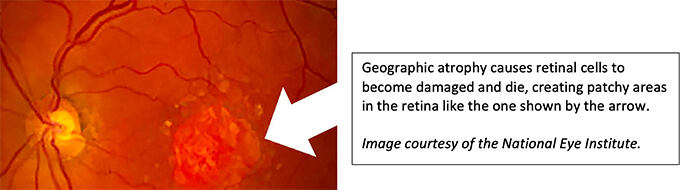
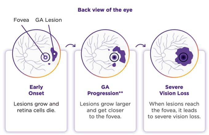
Geographic atrophy occurs when cells in the macula gradually break down and stop functioning. As these retinal cells are lost, areas of vision can become blurred, dim, or missing altogether.
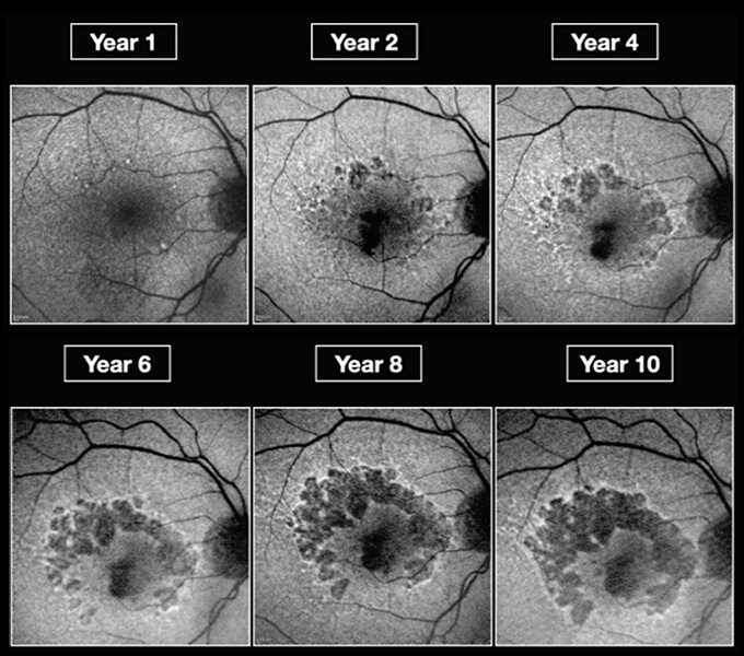
In the early stages of dry AMD, changes in the retina tend to be more subtle and may not cause noticeable vision loss immediately. Geographic atrophy represents a more advanced phase, in which damage to the macula becomes more pronounced and visible on retinal imaging. Over time, the affected areas can enlarge, leading to increasing difficulty with tasks that rely on central vision, such as reading, recognizing faces, or seeing fine details.
Geographic atrophy is a progressive condition, meaning it worsens over time. However, the speed of progression can vary significantly from person to person, which is why ongoing monitoring by a retina specialist is crucial.
Types of Geographic Atrophy
Geographic atrophy can present in different patterns within the macula. These patterns help retina specialists understand how the disease is affecting the retina and how it may change over time. Both types of geographic atrophy can progress and require regular follow-up to monitor vision changes and retinal health.
Focal Geographic Atrophy
Focal geographic atrophy involves a single, well-defined area of retinal cell loss in the macula. In the earlier stages, vision changes may be more localized, depending on where the atrophy is located. Some patients may notice a small blurred or missing spot in their central vision, while other areas remain relatively unaffected.
Multifocal Geographic Atrophy
Multifocal geographic atrophy occurs when multiple areas of retinal cell loss develop within the macula. Over time, these separate areas may expand and merge, leading to a broader impact on central vision. This pattern is often associated with more widespread visual changes and may affect daily activities sooner as the areas of atrophy increase.
Symptoms of Geographic Atrophy
The symptoms of geographic atrophy primarily affect central vision, which is responsible for seeing fine details. Because geographic atrophy develops gradually, changes may begin subtly and become more noticeable over time.
Common symptoms include:
- Central vision loss, which can make reading, recognizing faces, and seeing fine details more difficult
- Patchy or “missing” areas in the center of vision, where parts of words or objects may seem faded or absent
- Reduced contrast sensitivity, making it harder to distinguish objects from their background, especially in low light
- Increased difficulty with daily activities such as reading, driving, using screens, or recognizing familiar faces
It’s important to note that geographic atrophy typically does not affect peripheral (side) vision. Many patients retain their side vision even as central vision changes, which can help with mobility and orientation. Because symptoms can evolve over time, regular eye exams with a retina specialist are essential for tracking progression and adjusting care as needed.
Causes and Pathophysiology of Geographic Atrophy
Geographic atrophy develops as a result of gradual damage to retinal cells in the macula over time. These cells play a critical role in central vision, and once they are lost, they do not regenerate. The process is slow and progressive, which is why symptoms often worsen gradually rather than suddenly.
Several underlying biological processes are believed to contribute to the development of geographic atrophy. These may include:
- Oxidative stress, which can damage retinal cells as they age
- Chronic inflammation within the retina, leading to ongoing cellular injury
- Dysregulation of the complement system, a part of the body’s immune response that, when overactive, may contribute to retinal degeneration
Together, these processes can disrupt the health of the macula and accelerate the loss of retinal cells over time. While the exact cause of geographic atrophy is complex, ongoing research continues to improve understanding of how inflammation and retinal degeneration drive disease progression.
Risk factors:
Certain factors may increase a person’s risk of developing geographic atrophy, including:
- Advanced age
- Family history of macular degeneration
- Smoking, which increases oxidative stress in the retina
- Cardiovascular health conditions, such as high blood pressure or heart disease
- Long-standing dry age-related macular degeneration
Having one or more risk factors does not mean geographic atrophy is inevitable, but it does highlight the importance of regular eye exams and early monitoring.
Potential Complications of Geographic Atrophy
Because geographic atrophy affects the macula, its complications are primarily related to central vision. Over time, many patients experience progressive and irreversible loss of central vision, which can significantly affect daily life.
As areas of retinal cell loss enlarge, central scotomas (i.e., blind spots) may expand and become more noticeable. This can lead to increasing difficulty with everyday activities such as reading, driving, using digital devices, or recognizing faces. Some patients may also experience visual distortion (metamorphopsia), where straight lines appear wavy, or objects seem misshapen.
In some cases, patients with geographic atrophy may also develop wet age-related macular degeneration, a related but distinct condition that can cause more rapid vision changes and requires prompt treatment.
Beyond physical vision changes, geographic atrophy can have emotional and quality-of-life impacts. Frustration, anxiety, and a sense of loss of independence are common and important to address as part of comprehensive care.

How Geographic Atrophy Is Diagnosed
Geographic atrophy is diagnosed through a comprehensive eye exam and specialized retinal imaging performed by a retina specialist. Because early changes may not always be obvious to patients, detailed evaluation is essential for accurate diagnosis and ongoing care.
Diagnosis typically includes a dilated eye exam, which allows the retina specialist to closely examine the macula and surrounding retinal structures. Advanced imaging tools are also used to confirm the presence of geographic atrophy and track how it changes over time, including:
- Optical coherence tomography (OCT), which provides cross-sectional images of the retina and shows areas of retinal thinning and cell loss
- Fundus autofluorescence, which helps highlight areas of damaged or stressed retinal cells and can reveal patterns of atrophy
Because geographic atrophy is progressive, ongoing monitoring is critical. Regular follow-up visits allow retina specialists to detect changes early, measure progression, and adjust care as needed to protect remaining vision.
Treatment Options for Geographic Atrophy
At this time, geographic atrophy cannot be cured, and lost vision cannot be restored. However, treatment options are now available that may help slow the progression of the disease in some patients.
The FDA has approved medications specifically designed to slow geographic atrophy progression, including Syfovre® (pegcetacoplan) and Izervay® (avacincaptad pegol). These treatments work by targeting inflammatory pathways believed to play a role in retinal cell damage. Not every patient is a candidate, and treatment decisions are made carefully based on individual eye health and disease characteristics.
Living With Geographic Atrophy
Living with geographic atrophy can present daily challenges, but many patients find that supportive strategies help maintain independence and quality of life. Vision rehabilitation services and low-vision aids, such as magnifiers, enhanced lighting, and adaptive technology, can make everyday tasks more manageable.
Simple adjustments, like increasing contrast, using larger text, or modifying home lighting, can also help reduce visual strain. For many patients, learning new ways to approach familiar activities can restore confidence and functionality.
The emotional impact of vision changes is equally important to address. Feelings of frustration, anxiety, or grief are common, and emotional support or counseling may be helpful for some individuals. Staying engaged with ongoing care, attending regular follow-up appointments, and maintaining open communication with a retina specialist can provide reassurance and support throughout the course of the condition.
When to See a Retina Specialist and Why Ongoing Monitoring Matters
If you notice new or worsening changes in your central vision, such as difficulty reading, recognizing faces, or seeing fine details, it is important to see a retina specialist promptly. Changes on an Amsler grid, or concerns about progression in existing dry macular degeneration, should also be evaluated. Geographic atrophy requires specialized retina care and advanced imaging that goes beyond routine eye exams.
Because geographic atrophy progresses differently from person to person, ongoing monitoring plays a critical role in care. Regular visits allow a retina specialist to track changes in the retina, adjust treatment plans as needed, and detect early signs of conversion to wet macular degeneration. Building a long-term partnership with a retina specialist helps ensure timely intervention and continued support as vision needs evolve.
Frequently Asked Questions About Geographic Atrophy
Geographic atrophy is not the same as early macular degeneration, but it is an advanced form of dry age-related macular degeneration (AMD). In geographic atrophy, there is a significant loss of retinal cells in the macula, leading to more noticeable and progressive central vision changes than in earlier stages of dry AMD.
The progression of geographic atrophy varies from person to person. Some individuals experience slow changes over many years, while others notice more steady progression. Factors such as overall eye health and the pattern of atrophy can influence the rate of vision changes, which is why regular monitoring by a retina specialist is important.
Geographic atrophy itself does not involve bleeding or fluid, but some patients may develop wet macular degeneration in addition to geographic atrophy. Wet AMD can cause more rapid vision changes and requires prompt treatment, making regular eye exams essential for early detection.
Geographic atrophy often affects both eyes, though not always at the same time or to the same degree. One eye may show more advanced changes than the other. Because progression can differ between eyes, each eye is monitored individually during follow-up visits.
Schedule a Consultation for Geographic Atrophy in South Carolina
Geographic atrophy is a progressive condition, but early diagnosis, specialized care, and ongoing monitoring can play an important role in protecting your remaining vision. Working with a retina specialist ensures access to advanced imaging, personalized treatment options, and long-term support.
If you have been diagnosed with geographic atrophy or are experiencing changes in your central vision, contact Retina Consultants of South Carolina to schedule a consultation with our retina specialists.